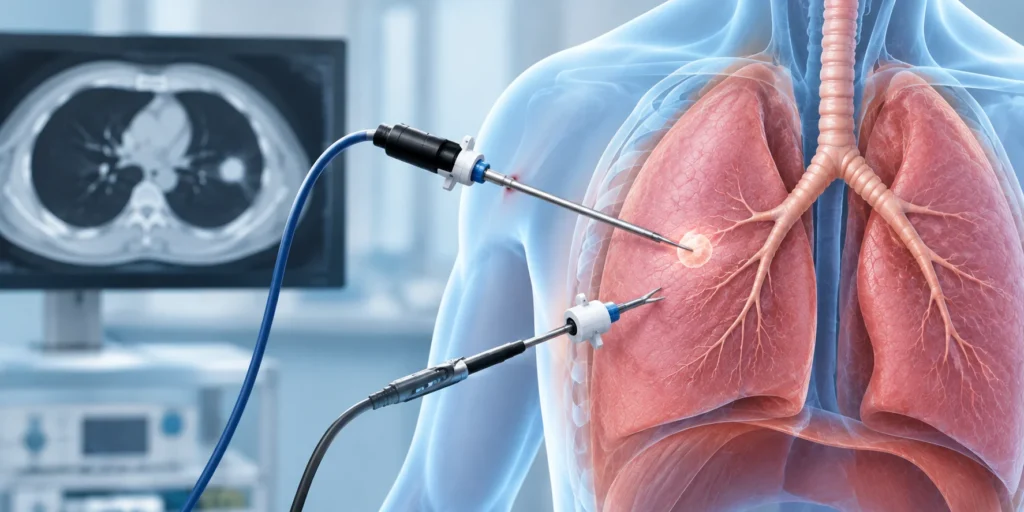
A nodule or shadow on a lung scan is rarely explained by the scan itself. Getting tissue is the only way to know what’s actually there. A VATS lung biopsy takes a small sample directly from the lung using a camera and instruments passed through keyhole cuts in the chest wall, no large incision, no spreading the ribs. Something that once meant weeks of recovery now means a hospital stay of a day or two.
According to Dr. George Karimundackal, a leadingThoracic Surgeon in Mumbai, “A lot of patients are sent for a lung biopsy not knowing which route their surgeon will take. The VATS approach gives us a clear view inside the chest and a proper tissue sample in one sitting. We’re not guessing from a needle through the skin. We see exactly where the nodule is, and we take exactly what the pathologist needs.”
How Does a VATS Lung Biopsy Actually Work?
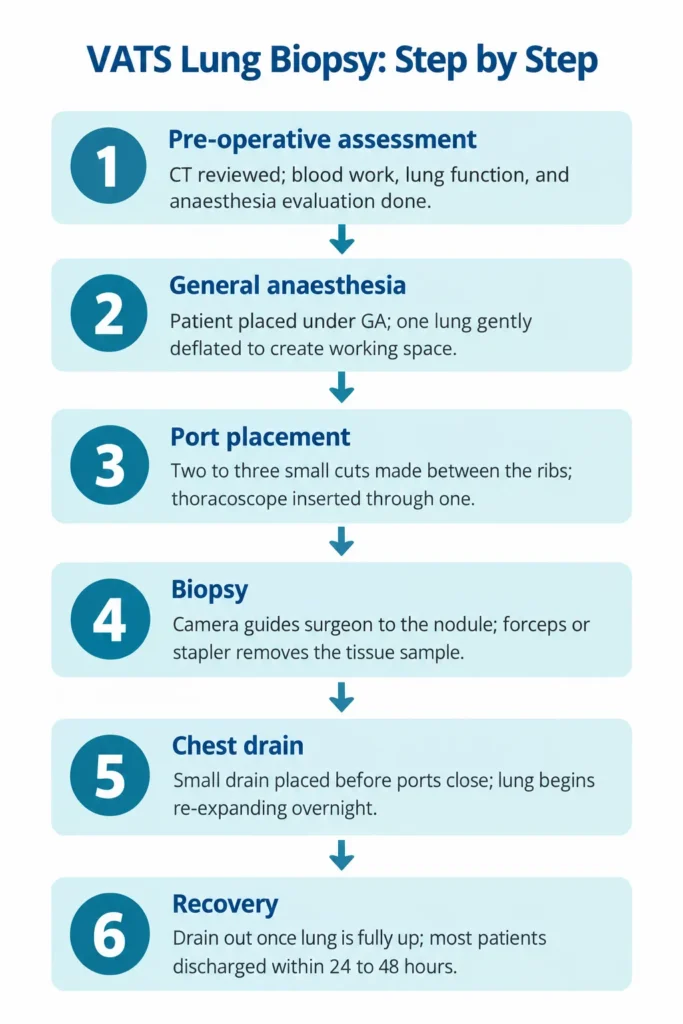
The procedure runs in steps, each one setting up the next. The infographic below maps the full sequence from prep to discharge.
What the steps don’t capture is the work that happens before step one. Every nodule’s exact position, its proximity to vessels and the chest wall, gets planned on CT beforehand. That mapping is what keeps the biopsy itself quick and precise, not exploratory. The full range of what VATS can do beyond a biopsy is covered on theVATS surgery page.
How Does VATS Biopsy Compare to Other Lung Biopsy Methods?
Not every nodule gets biopsied the same way. Where it sits and what the result needs to rule out usually settles the choice.
Feature
VATS Lung Biopsy
CT-Guided Needle Biopsy
Bronchoscopic Biopsy
Access route
Keyhole surgery through chest wall
Needle through skin under CT
Camera through airway
Best for
Peripheral or larger nodules needing definitive diagnosis
Peripheral nodules accessible by needle
Central lesions near airways
Sample quality
Larger, more representative tissue sample
Small core; may be insufficient
Small brushing or washing
Pneumothorax risk
Low, drain placed at surgery
Up to 15-20%
Very low
Anaesthesia
General
Local or sedation
Sedation
Hospital stay
1 to 2 days
Day procedure
Day procedure
Sample size is what changes everything: VATS pulls a full wedge of tissue. That wedge gives the pathologist enough material to confirm tumour type, grade, and molecular markers. A needle core often can’t.
Where the nodule sits decides the tool: peripheral lesions tucked close to the chest wall suit VATS well. Lesions near the bronchi get a bronchoscope first, since accessing them through the wall doesn’t make sense.
One visit, one answer: small suspicious nodules can be fully excised during the same VATS biopsy. If the result comes back malignant and the lesion was small, no second procedure is needed.
Drain before you close: a needle biopsy can cause a pneumothorax that shows up hours later without warning. With VATS, the drain goes in before the ports close and the lung re-expansion is watched from the start.
Because VATS covers diagnosis and potential resection in a single sitting, understanding the risks it carries matters too. Our post onVATS surgery risks covers those in full.
Why Choose Dr. George Karimundackal?
Dr. George Karimundackal is one of India’s leading thoracic surgeons with over 15 years of experience. He was previously Professor of Thoracic Surgery at Tata Memorial Hospital in Mumbai before taking up his current role as Director, Thoracic Surgery at Nanavati Max Hospital. Lung biopsy and wedge resection are a routine part of his practice, with over 1,000 VATS procedures performed. Before any biopsy is planned, he reviews the CT in detail to confirm whether VATS, a needle approach, or another route actually suits the lesion.
Frequently Asked Questions
What is a VATS lung biopsy?
A minimally invasive procedure using a camera to remove a small lung tissue sample.
How long does a VATS biopsy take?
Usually 30 to 60 minutes, depending on the size and location of the nodule.
Is a VATS biopsy painful?
General anaesthesia is used; post-operative soreness is mild and short-lived.
How soon are biopsy results available?
Most histology results are available within five to seven working days.
Disclaimer: This blog is for informational purposes only and isn’t a substitute for professional medical advice; please consult a specialist for diagnosis and treatment.