
Pneumothorax Treatment Guidelines
A pneumothorax, commonly referred to as a collapsed lung, occurs when air leaks into the pleural space and causes the lung to partially or fully collapse. It can develop spontaneously, follow a chest injury, or arise from an underlying lung condition. The treatment approach, whether observation, aspiration, chest tube drainage, or surgery, depends on several factors and the clinical presentation of each case.
“The treatment path for a pneumothorax is rarely one-size-fits-all,” says Dr. George Karimundackal, a highly regarded thoracic surgeon in Mumbai, India. “The size of the collapse, the type, and whether it has happened before all determine what we do next. Getting that assessment right from the start is what prevents recurrence and avoids unnecessary procedures.”
With over 15 years of experience, Dr. Karimundackal specializes in managing primary, secondary, and recurrent pneumothorax cases. His expertise covers the full range of collapsed lung treatment, including aspiration, chest tube management, and VATS surgery. For patients seeking clarity on pneumothorax treatment guidelines, Dr. Karimundackal brings a structured, evidence-based approach grounded in high-volume thoracic practice at one of India’s foremost thoracic surgery centres.
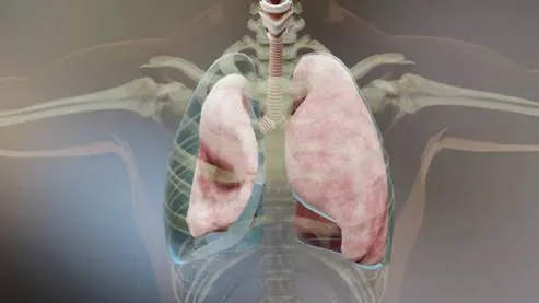
Not everyone arrives at a diagnosis knowing what a pneumothorax actually is. Let’s explore the condition before getting into how it is treated.
What Is Pneumothorax?
A pneumothorax occurs when air leaks into the pleural space, the gap between the lung and chest wall, causing the lung to partially or fully collapse. When air enters this space, it pushes against the lung and prevents it from expanding properly.
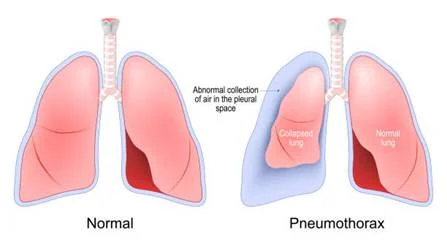

There are three main types. A primary spontaneous pneumothorax affects otherwise healthy individuals, usually due to ruptured air sacs called blebs. A secondary spontaneous pneumothorax develops in patients with existing lung disease like COPD or asthma, and carries a higher risk. A traumatic pneumothorax results from a chest injury or medical procedure. A tension pneumothorax is a life-threatening emergency requiring immediate collapsed lung treatment.
Identifying the type is the starting point of pneumothorax management and directly shapes the treatment approach.
What factors go into choosing between observation, aspiration, a chest tube, or surgery? Let’s delve into the clinical decision-making process behind pneumothorax management.
How Do Doctors Decide the Right Treatment for Pneumothorax?
No two pneumothorax cases are identical, and treatment decisions depend on several factors assessed together:
Size of the pneumothorax:
A small pneumothorax, typically less than 2 cm on a chest X-ray, may be managed conservatively. A large one almost always needs active intervention.
Type:
Primary spontaneous cases in otherwise healthy patients have more conservative treatment options than secondary cases in patients with compromised lung function.
Symptoms:
A patient who is breathless at rest needs faster intervention than one who is only mildly uncomfortable.
First episode vs. recurrence:
A second or third pneumothorax on the same side significantly changes the treatment conversation, often bringing VATS surgery into the picture.
Occupation and lifestyle:
Divers and pilots face specific restrictions and may be advised to undergo surgical treatment even after a first episode due to the risk of recurrence in their professions.
Underlying lung disease:
Secondary pneumothorax in patients with reduced lung reserve requires more urgent intervention and closer monitoring.
Early expert assessment can prevent a manageable situation from becoming serious. Get in touch with a thoracic specialist for a personalised evaluation today.
Which patients qualify for needle aspiration as a first step? Let’s discover when this minimally invasive option is appropriate.
When Is Needle Aspiration Recommended?
Needle aspiration, also called simple aspiration, involves inserting a thin needle into the pleural space and drawing out the trapped air with a syringe. It is done under local anaesthesia, typically takes 15 to 30 minutes, and does not require a hospital stay in many cases.
Current spontaneous pneumothorax treatment guidelines recommend needle aspiration as a first-line option for:
- Primary spontaneous pneumothorax with a large air gap (greater than 2 cm) but in a patient who is clinically stable and not significantly breathless
- First-episode pneumothorax in young, otherwise healthy patients
Aspiration works in roughly 50 to 70 percent of primary spontaneous cases. If the lung re-expands fully on a follow-up X-ray taken two to four hours later, the patient can often be discharged with instructions to return if symptoms worsen. If aspiration fails or the lung does not fully expand, the next step is chest tube insertion.
Aspiration is generally not suitable for secondary pneumothorax or for patients who are haemodynamically unstable.

When does collapsed lung treatment move beyond aspiration? Let’s dive into when a chest tube becomes necessary.
When Is a Chest Tube Needed?

A chest tube, also called an intercostal drain, is a small flexible tube inserted between the ribs into the pleural space to continuously drain air and allow the lung to re-expand. It is connected to an underwater seal drainage system or a one-way valve that prevents air from re-entering.
Failed needle aspiration, where the lung has not re-expanded after aspiration
Secondary spontaneous pneumothorax, where conservative management carries a higher risk
Large or symptomatic pneumothorax in patients who are breathless or haemodynamically compromised
Traumatic pneumothorax, including haemopneumothorax (blood and air in the pleural space)
Tension pneumothorax, where emergency decompression is needed
Small-bore chest tubes (10-14 Fr) are now preferred over larger ones in most cases, as they are equally effective, cause less discomfort, and are associated with fewer complications. The tube typically stays in place for two to five days, until the air leak resolves and the lung remains expanded on clamping.
If the air leak persists beyond five to seven days or if the lung fails to fully expand, surgical review is recommended.
At what point does pneumothorax management move beyond tubes and aspiration into surgical territory? Let’s discuss the clinical situations where surgery becomes the right path forward.
When Is Surgery Recommended for Pneumothorax?
Surgery for pneumothorax is not a last resort. In certain situations, it is the most appropriate and definitive treatment available. Current guidelines recommend surgical intervention for:
Recurrent ipsilateral pneumothorax (second or subsequent occurrence on the same side)
Persistent air leak lasting more than five to seven days despite chest tube drainage
Failure of the lung to re-expand after chest tube placement
Bilateral pneumothorax, where both lungs are affected, either simultaneously or sequentially
First-episode pneumothorax in high-risk professions, such as pilots and divers, where recurrence carries life-threatening consequences

Haemothorax (bleeding into the pleural space) alongside the pneumothorax
Underlying blebs or bullae identified on the CT scan, which are likely to rupture again
“The surgical goal is twofold: to remove the blebs or damaged tissue causing the air leak, and to create adhesions between the lung surface and chest wall (pleurodesis) so that a future pneumothorax is far less likely,” explains Dr. George Karimundackal.
The right surgical decision can change your long-term outcome. Connect with a thoracic specialist to find out if surgery is the right step for you.
With more than one surgical technique available, why has VATS emerged as the standard surgical choice for pneumothorax? Let’s explore the reasons behind this shift.
Why Is VATS Considered the Preferred Surgical Option?
Open thoracotomy was once the only surgical route for pneumothorax. It involved a large chest incision, significant post-operative pain, and a recovery of six weeks or more. VATS (Video-Assisted Thoracoscopic Surgery) has largely replaced it for most pneumothorax cases, and the reasons are well-supported by evidence.

During VATS for pneumothorax, the surgeon makes two to three small incisions and uses a thoracoscope to visualise the lung surface. Blebs are identified and removed (bullectomy), and pleurodesis is performed either mechanically by abrading the pleural surface or by applying a chemical agent like talc.
The advantages over open surgery include:
- Shorter hospital stay, typically two to three days
- Significantly less post-operative pain
- Faster return to work and normal activity
- Lower risk of wound infection
- Equally effective recurrence prevention when performed correctly
Recurrence rates after VATS pleurodesis are reported at less than five percent, which is comparable to open surgery. For patients with a second pneumothorax or a persistent air leak, VATS is now the internationally recommended surgical approach across most thoracic surgery guidelines.
What needs to change in daily life once the lung has healed? Let’s delve into the precautions that reduce the risk of recurrence.
Lifestyle Precautions After a Pneumothorax
Whether treated conservatively or surgically, lifestyle adjustments matter in the weeks and months after a pneumothorax:
- Avoid smoking completely: Smoking is a major risk factor for bleb formation and recurrence. Stopping smoking is the single most impactful lifestyle change a patient can make.
- No air travel for at least two weeks after full lung re-expansion has been confirmed on imaging. Cabin pressure changes can trigger a recurrence.
- Avoid scuba diving permanently if you have had a pneumothorax and have not had surgical pleurodesis. Even after surgery, diving clearance requires specialist review.
- Avoid heavy lifting and strenuous exercise for at least two to four weeks post-treatment, or longer after surgery.
- Watch for symptoms: Any return of chest pain, breathlessness, or a feeling of tightness warrants an immediate medical review, even after a confirmed recovery.
- Attend all follow-up appointments: A repeat chest X-ray at two to four weeks confirms that the lung remains fully expanded.

Dr. George Karimundackal advises, “Patients treated for secondary pneumothorax should continue management of their underlying lung condition closely, as poorly controlled COPD or asthma significantly raises the risk of another episode.”
Some symptoms after a pneumothorax, or even before a diagnosis, signal an emergency. Let’s discover the warning signs that require an immediate visit to the emergency department.
When Should You Seek Immediate Medical Attention?

Seek emergency medical care without delay if you or someone around you experiences:
Sudden, severe breathlessness that comes on rapidly and does not ease
Sharp, stabbing chest pain on one side, particularly after physical activity or a coughing fit
Rapid heart rate combined with breathlessness and a feeling of lightheadedness
Bluish discolouration of the lips or fingertips (cyanosis), which suggests the body is not getting enough oxygen
A drop in blood pressure or feeling faint, which may indicate a tension pneumothorax
Breathlessness returning after a period of confirmed recovery, as this may signal a recurrence
A tension pneumothorax, in particular, can deteriorate within minutes. Do not wait to see if symptoms improve on their own. Call emergency services or go directly to the nearest hospital.
Frequently Asked Questions
A small primary pneumothorax in an otherwise healthy, stable patient can resolve without intervention as the body reabsorbs the trapped air. Larger or secondary cases always require active treatment.
It depends on the size, type, and whether it is a first or recurrent episode. Options range from observation and needle aspiration to chest tube drainage or VATS surgery for complex or recurring cases.
Conservative cases recover within one to two weeks. Chest tube patients typically recover in two to three weeks. After VATS surgery, most patients return to normal activity within four to six weeks.
Recurrence rates after a first episode managed without surgery range from 25 to 50 percent. VATS surgery with pleurodesis reduces this risk to below five percent.
Yes. It most commonly affects tall, thin young men between 15 and 35 years. Smokers and patients with underlying lung conditions like COPD face a significantly higher risk.