Esophageal Cancer Doctor in Mumbai
Esophageal cancer treatment is complex and requires a dedicated team of specialists including surgeons, oncologists, nutritionists, physiotherapists and many others. Dr. George Karimundackal is an expert thoracic surgeon in Mumbai with significant experience in esophageal surgery. He is an expert in VATS and robotic (RATS) esophagectomy and has conducted many training programs in these techniques. He is also an expert in complex esophageal surgeries for corrosive injuries which may require the replacement of the esophagus with the large bowel(coloplasty). Dr. George is ably supported in these complex procedures by a dedicated team of specialists who provide comprehensive care.
About Esophageal Cancer
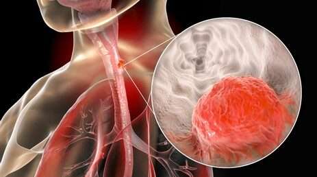
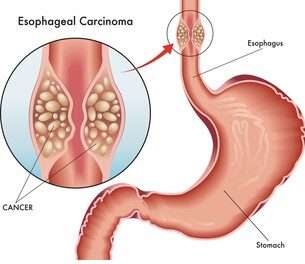
The exact cause of esophageal cancer is not yet fully understood. Esophageal cancer may be associated with chronic irritation of the esophagus, smoking, heavy alcohol consumption, gastroesophageal reflux disease (GERD) and frequent consumption of very hot liquids
The symptoms of esophageal cancer
Unfortunately, early-stage esophageal cancer does not show many signs or symptoms and patients rarely present in this stage. As the disease progresses and obstructs the food pipe, or spreads to adjacent lymph nodes patients develop symptoms. The symptoms may be any or all of the following
- Difficulty in swallowing or pain while swallowing
- Loss of weight
- Vomiting or regurgitation immediately after having food
- Alteration of voice
- Cough after consuming food
- Backache
It is recommended that you consult a medical expert at the earliest if you have any of these symptoms. An early diagnosis significantly affects treatment and outcomes.
Diagnosis of Esophageal Cancer
The diagnosis of esophageal cancer usually begins with a detailed medical history, physical examination followed by a number of tests to confirm esophageal cancer and to stage it.


Stages of esophageal cancer and treatment strategy
Esophageal cancer staging like in any other cancer depends on the extent of the primary disease in the esophagus (T stage), spread to adjacent lymph nodes (N stage) and spread to distant organs (M stage). For ease of understanding and simplifying treatment treatment strategy it is easier to classify esophageal cancer into early stage disease, locally advanced disease and metastatic disease (stage 4).
The choice of treatment in esophageal cancer depends on not only the stage of disease but also on the location of the tumour and the fitness of the patient to tolerate treatment. The decision is usually taken by the primary physician in consultation with other specialists so that the patient gets a comprehensive treatment plan.
- Early stage esophageal cancer is disease limited to the esophageal mucosa or submucosa with no lymph nodal involvement. Such disease can be treated with endoscopic removal or surgical removal of the esophagus. A proper assessment by endoscopic ultrasound (EUS) is mandatory before starting treatment. In disease involving the upper third of the esophagus radical radiotherapy may be the best option for treatment. No adjuvant treatment is required and survival rates are excellent.
- Locally advanced disease includes more advanced primary disease in the esophagus with spread to adjacent lymph nodes. These patients need multimodality management including surgery, chemotherapy and radiotherapy, and the results are promising. Disease involving the upper third of the esophagus are generally treated with chemotherapy and radiotherapy together. Diseases of the middle and lower third of the esophagus are better treated with surgery. However these patients need to be pretreated before surgery. In patients with squamous cell cancer chemoradiotherapy is preferred before surgery, while in patients with adenocarcinoma, only chemotherapy is required along with surgery.
In patients with metastatic esophageal cancer, the focus shifts from attempting cure to relieving symptoms. The main symptoms are difficulty in swallowing and occasionally aspiration. The usual strategies for relieving swallowing issues are radiotherapy and placement of esophageal stents. Both modalities have their benefits and the choice of modality is taken on a case to case basis. Systemic therapy in the form of chemotherapy is also useful and may help in reducing tumour bulk and extend life.
Endoscopic mucosal resection:
Endoscopic mucosal resection is a minimally invasive procedure performed to remove the precancerous or cancerous tissue or abnormal tissues from the lining of the esophagus using an endoscope. The endoscope is a long, narrow tube fitted with a light camera and other instruments. It is passed through the throat and can access the esophagus and the stomach. This is suitable only for very early stage tumours affecting only the mucosa
Surgery
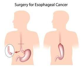
- Transthoracic esophagectomy : It is the most common type of esophageal cancer surgery. It is performed through an incision in the chest, abdomen and the neck. The surgeon removes most of the esophagus and a small part of the stomach, then joins the stomach to the remaining esophagus in the neck.
- Ivor Lewis Esophagectomy: Ivor Lewis esophagectomy is also a very common type of esophagectomy in which the surgeon makes incisions in the chest and the abdomen. The surgeon will remove a part of the esophagus, make a tube using some part of the stomach and then connect it to the esophagus in the chest.
- Thoracoabdominal esophagogastrectomy: The thoracoabdominal approach is a special procedure to remove a tumor detected in the lower esophagus or gastroesophageal (GE) junction. In this procedure, the surgeon makes an incision the left side, i.e. the chest and the abdomen and divides the esophagus in the left chest. The continuity of the gut is restored using the stomach tube or the small intestine by connecting it to the native esohphagus in the chest.
Post operative recovery
The patient recover gradually over a few days and the usual time to discharge is around 9 to 10 days. Patients are started on regular chest physiotherapy and tube feeds at the earliest. This is continued till the the newly formed connections in the bowel heal, which is usually around the 6th or 7th day. At the time of discharge patients will be on full oral feeds and active enough to carry out most activities of routine life.Frequently Asked Questions
The usual hospital stay is around 9 to 10 days and patients are able to get back to light work by day 21. After esophagectomy, surgeons recommend consuming smaller, more frequent meals and sleeping with the head end of the bed slightly elevated. Also patients are advised to take life long vitamin supplements.
Esophageal cancer can sometimes be cured if diagnosed and treated early. However, the success of the treatment depends on the cancer stage, the type of treatment used, and the patient’s overall health.
More than 15 out of every 100 cases (more than 15%), cancer patients can live for at least five years after diagnosis. More than ten people in 100 (10%) will live with their cancer for ten years or more.
Some stage 4 esophageal cancer patients may live for more than a year. Around 20 out of 100 patients (or 20%) with stage 4 esophageal cancer will still be alive a year after diagnosis


