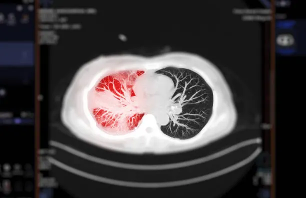
Hearing that your CT scan shows a Ground Glass Nodule (GGO) on your CT scan can be worrying. Many people immediately fear lung cancer, but the reality is far more reassuring. Ground glass nodules are common findings on modern CT scans, especially because imaging technology has become much more sensitive. While some GGOs may represent early lung cancer, many are caused by temporary inflammation, infections, or benign lung conditions that disappear on their own.
According to Dr. George Karimundackal, careful evaluation is the key to managing GGOs. “Not every ground glass nodule requires surgery. The size, appearance, growth pattern, and patient history help determine whether observation or treatment is the right approach. The goal is to treat patients at the right time while avoiding unnecessary surgery.”
As a leading lung cancer specialist & thoracic surgeon in Mumbai, Dr. George Karimundackal specializes in evaluating lung nodules, performing minimally invasive lung surgery, and treating early lung cancers using advanced thoracic techniques. His patient centered approach ensures every case receives individualized care based on the latest international guidelines.
Confused about what a ground glass nodule on your CT scan means? Let’s break down this finding in simple terms.
What Exactly is a Ground Glass Nodule?
A ground glass nodule (GGO) is a hazy area seen on a CT scan that appears lighter than normal lung tissue but does not completely block the underlying lung structures. Unlike a solid lung nodule, blood vessels and airways can still be seen through a GGO.
A ground glass nodule is not a diagnosis. Instead, it is a radiological finding that may have several possible causes.
Common causes include:
Lung infections
Viral pneumonia
Inflammation
Pulmonary fibrosis
Organizing pneumonia
Early lung adenocarcinoma
Pre-cancerous lung lesions
Many GGOs disappear after treating an infection, while others remain stable for years without causing problems.
Not all GGOs behave the same way. Let’s compare the two main types doctors look for.
Pure GGO vs Part Solid Nodule: Why the Difference Matters
Pure and part-solid nodules differ in appearance, cancer risk, and follow-up needs. Here is a quick comparison:
Feature
Pure Ground Glass Nodule
Part Solid Nodule
Appearance
Completely hazy
Hazy with a solid component
Cancer Risk
Lower
Higher
Growth Rate
Usually slow
Often faster
Follow up
Usually CT surveillance
Closer monitoring
Surgery
Only if persistent or enlarging
More likely if solid part grows
Prognosis
Excellent when treated
Excellent with early surgery
The presence of a solid component is one of the most important predictors of malignancy.
This is the question most patients ask first. Let’s separate facts from fear.
Does a Ground Glass Nodule Mean Cancer?
No. Most GGOs are not cancer.
The likelihood depends on several factors:
Size
Shape
Patient’s age
Smoking history
Family history
Growth over time
Presence of a solid component
Persistent GGOs that slowly enlarge over several years deserve closer evaluation because they may represent very early lung adenocarcinoma.
The good news is that when detected at this stage, treatment outcomes are excellent.
Patients with early lung cancer diagnosed through CT screening often achieve cure rates above 90 percent after surgery.
Every small detail on the CT scan matters. Let’s explore what specialists carefully evaluate.
What the Size and Features Tell your Surgeon
A thoracic surgeon looks beyond simply measuring the nodule.
Important features include:
Size
Less than 6 mm
6 to 10 mm
Larger than 10 mm
Larger nodules generally require closer surveillance.
Shape
Smooth borders are generally more reassuring than irregular or spiculated margins.
Density
A persistent solid component increases suspicion.
Growth
Even slow enlargement over several years can be significant.
Number of Nodules
Multiple GGOs may indicate inflammation or certain types of lung disease.
Patient Factors
Your surgeon also considers:
Smoking history
Previous cancers
Family history
Occupational exposure
Lung function
This complete assessment helps determine whether continued observation or intervention is appropriate.
Why doctor recommends another CT instead of surgery? Let’s understand why monitoring is often the safest option.
The Follow-up Timeline: Why "just watch it" is the Right Call
Many patients become anxious when told to “watch and wait.” In reality, careful surveillance is supported by international guidelines.
Typical follow-up may include:
Repeat CT after 3 months if infection is suspected
CT after 6 to 12 months for persistent small GGOs
Annual CT scans for stable nodules
Longer surveillance for selected patients
The purpose is to identify any change in:
Size
Density
Shape
Development of a solid component
Most stable GGOs never require surgery.
Avoiding unnecessary operations is just as important as treating cancer early.
Some nodules eventually require treatment. Let’s see when surgery becomes the best option.
When is Surgery Actually Needed for a GGO?
Surgery may be recommended when:
The nodule continues growing
A solid component develops
Cancer is strongly suspected
PET scan or biopsy supports malignancy
Patient preference after detailed discussion
Whenever possible, minimally invasive surgery is preferred.
Not every lung nodule needs surgery, but every persistent nodule deserves expert review. Let’s understand why specialist evaluation makes a difference.
Why a Thoracic Surgeon Should Review your GGO
A thoracic surgeon understands both surgical and non-surgical management.
Benefits of consulting an experienced specialist include:
Accurate interpretation of CT findings
Review of previous imaging for comparison
Assessment of cancer risk
Guidance on surveillance schedules
Minimally invasive treatment when required
Multidisciplinary care with pulmonologists, radiologists, and oncologists
As an experienced lung cancer specialist in Mumbai, Dr. George Karimundackal specializes in evaluating complex lung nodules and treating early lung cancer using advanced minimally invasive techniques.
Frequently Asked Questions
What is a ground glass nodule in the lung?
It is a hazy area seen on a CT scan that may represent inflammation, infection, scarring, or an early lung tumor.
Does every ground glass nodule become cancer?
No. Most GGOs remain benign or stable and never become cancerous.
What size ground glass nodule is concerning?
Persistent nodules larger than 10 mm or those developing a solid component require closer evaluation.
Can non-smokers develop ground glass nodules?
Yes. Many GGOs occur in non-smokers due to infections, inflammation, or early lung adenocarcinoma.
Is VATS surgery used for ground glass nodules?
Yes. Video Assisted Thoracoscopic Surgery is commonly used to remove suspicious or cancerous lung nodules with minimal invasion.
What symptoms do ground glass nodules cause?
Most GGOs cause no symptoms and are discovered incidentally during CT scans.
Can ground glass nodules recur after surgery?
Recurrence is uncommon after complete removal of early-stage lesions, but regular follow-up remains important.